


Victor Nozais
Neuroimaging the white matter function in the living human brain
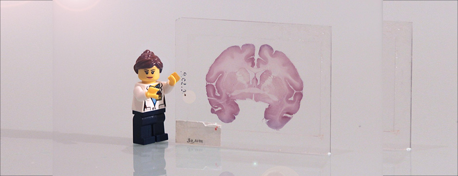
mars 2023 Directeur(s) de thèse : Résumé de thèseThis PhD project is focused on the functional mapping of the white matter of the human brain, which is relatively unexplored at the moment. Functional mapping consists in associating cognitive functions with their neuronal substrates, thus obtaining a better understanding of the relationships between structure and function organising the brain. However, functional mapping of the brain has mainly been focused on the study of grey matter in human neuroimaging. This bias comes from limitations in the methods, in particular magnetic resonance imaging (MRI), that results in a conceptual bias, a vision of cognitive networks limited to grey matter. And although the majority of synapses are indeed concentrated in grey matter, ignoring axon-mediated brain connectivity (in white matter) when studying brain function limits our understanding of the interaction between brain regions and the emergence of cognitive functions.
To enable the community to overcome these conceptual and technical barriers, we focused the first study of the thesis on the development of a method, the Functionnectome, that combines functional and structural connectivity information from MRI to offer a more integrated view of the brain and allow cognitive circuits to be represented directly on the white matter.
In the second study, we focused on characterising the brain’s functional organisation on a global scale in both grey and white matter. For this purpose, we used the “resting-state” paradigm in functional MRI (fMRI), i.e. the study of spontaneous fluctuations in the brain’s functional signal outside of a specific cognitive task (and therefore, at rest). These fluctuations generally reveal resting-state networks, which can then be used to functionally characterise the entire grey matter. In our study, we used the Functionnectome to combine the classical resting-state signal with white matter connectivity information, and thus study resting-state networks directly on white matter. We thus created WhiteRest, the first comprehensive atlas of resting-state networks showing both their grey and white matter coverage. We then validated WhiteRest by associating some of these networks with brain lesions in the white matter, demonstrating a match between symptoms and disruption of the studied networks.
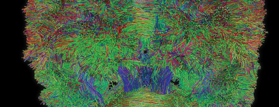
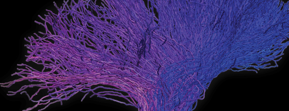
Finally, in the third study, we focused on improving the structural connectivity data we provide with the Functionnectome. These data, generated by tractography, were better optimised for the structural-functional analysis of the Functionnectome. First, we improved the interface between grey matter and white matter fibres, allowing better integration of the two types of information. Second, we divided the fibres according to their type of connectivity (association, projection, or commissural), which reduced some of the negative effects of fibre crossing in the white matter, and facilitated the interpretation of the functional maps generated by the Functionnectome.
In conclusion, through the Functionnectome, we have created a new technical and conceptual framework to reintegrate white matter at the centre of our understanding of cognitive networks in the healthy brain. We hope that the demonstration of its effectiveness will encourage the community to pursue and extend this new approach to the functional study of the brain.